More accurate gilt testing needed to detect Mycoplasma hyopneumoniae


Listen to the podcast


Sow herds seeking negative Mycoplasma hyopneumoniae (M. hyo) status should use accurate gilt surveillance methods.

Research conducted by Alyssa Betlach, DVM, veterinarian with Swine Vet Center and graduate student at the University of Minnesota, showed differences in the ability to detect a recent M. hyo infection based on sample types used.

“My research is focused on replacement gilt populations and really ensuring, if there was a recent introduction of M. hyo, were we able to detect such introduction with the commonly utilized practices we do today,” Betlach explained.

Maintaining a negative health status for M. hyo in the sow herd is important for reducing the economic toll that this pathogen has on hog production.

Detection differences based on sample type

In the research trial, a naturally infected gilt was introduced into a naïve population. The group was tested periodically for M. hyo using blood samples, oral fluids and other sampling methods including laryngeal swabs and deep tracheal catheters.

“There was one new naïve gilt that became positive at 6 weeks after contact, so you can appreciate that there’s a very slow transmission of M. hyo compared to other pathogens,” Betlach said.

The blood tests did not detect the naturally infected gilt until week 6. But the laryngeal swabs and deep tracheal catheters detected the infected gilt from the beginning of the trial. Oral fluids remained negative.

Different mindset required

The laryngeal swabs and deep tracheal catheters are relatively new sampling techniques. Both require training and practice but are more accurate than other sample types during the early stage of infection.

“If we really want to increase our ability to track M. hyo, we need to have a different mindset and focus in on more sample types that are more sensitive for detecting such pathogens.”

Producers should also understand the advantages and limitations of each sample type and make adjustments as needed in gilt surveillance.

“You could think about even extending your isolation period if you want to take a different sample or less sample sizes,” Betlach suggested.  

The end goal is achieving and maintaining a negative health status in sow herds.

Gilts and sows have a critical role in the transmission of M. hyo. “So it’s really important to minimize the introduction of it…to reduce the cost of M. hyo,” she said.


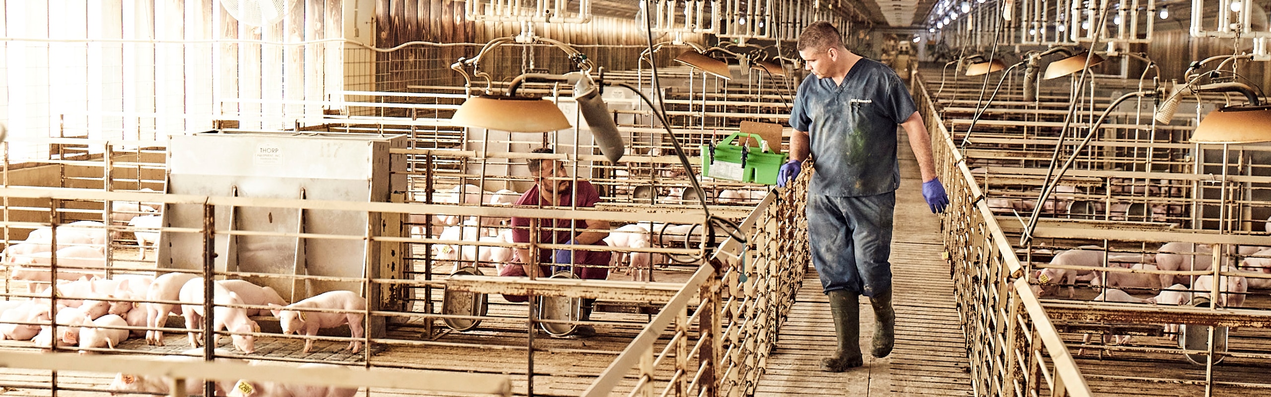
Gilt acclimatization is key to eliminating Mycoplasma hyopneumoniae in sow herd

Successful elimination of Mycoplasma hyopneumoniae (M. hyo) from a herd is often driven by sow farm status, according to Alyssa Betlach, DVM, Swine Vet Center. Betlach has researched M. hyo for several years in a PhD program at the University of Minnesota.

Within the industry, M. hyo continues to be a prevalent and economically important respiratory pathogen worldwide. Studies have shown that the disease can add $3 to $10 per pig due to decreased animal performance, including longer time to market and increased antimicrobial usage, she added.

Traits of M. hyo

Three unique features of M. hyo must be considered when developing a control plan. First, piglets are not born with M. hyo but often are colonized from their mothers, who shed it during farrowing. The bacterium colonizes in the lungs of the pigs and causes clinical signs in grow-finish due to the slow, chronic nature of the infection.

Second, M. hyo primarily resides in the respiratory tract by adhering to lung cilia.  Therefore, “When you think of diagnostics and sampling, you need to consider that it resides deep in the lung,” Betlach said.

Third, shedding from M. hyo has been shown to last up to 240 days and transmission is very slow. One pig needs 4 to 6 weeks to transmit the disease to another pig versus the flu that is transmitted from one pig to 14 others in a 2-week period.

Elimination starts on sow farm

Current elimination programs have successfully removed M. hyo from many herds. But the methods for elimination continue to improve by focusing on gilt acclimatization and sow farm stability.

“We need to think about negative gilts,” Betlach said. “If you have a positive farm and you constantly bring in negative gilts, you are adding wood to that fire. Therefore, we need to think of ways to address this.

“Gilt acclimatization is something we should think about,” she continued. “Gilt acclimatization strategies are designed to stabilize the sow herd by promoting ways to minimize shedding at farrowing.”

Acclimatizing gilts

Three types of gilt acclimatization are used, namely vaccination, natural exposure and exposure using a herd-specific lung homogenate. Vaccination for M. hyo is commonly practiced as it can reduce the bacterial load and clinical severity. However, M. hyo vaccination does not prevent colonization and has minimal effect on altering transmission.

Natural exposure through the introduction of M. hyo-positive culls into a gilt development unit has been performed. However, this approach is less than ideal in larger herds, as it takes a long time for adequate exposure and requires a high number of positive culls. Studies have shown that it takes one positive cull to one or two gilts for successful exposure within a 4-week period.

Exposure using a herd-specific, M. hyo-positive lung homogenate has been performed in the field, via intra-tracheal or aerosol techniques. Intra-tracheal exposure is a more labor-intensive method and may not be as feasible in large populations, as approximately 50% to 70% of the herd will need to be exposed to be successful. Therefore, aerosol exposure using foggers has been explored. Recently, this method has been shown to be successful for M. hyo exposure while being more feasible to perform.

Diagnostics for M. hyo

Several sample types are used for the detection of M. hyo. It is important to consider the overall goal that you are trying to achieve, as this will determine what sampling method should be used.

“Serum is commonly used, and the cost is relatively low,” Betlach said. “This sample type is used to detect the presence of M. hyo antibodies and it is easy to collect. However, the ability to interpret diagnostic results can be difficult, especially in vaccinated herds as the presence of antibodies from the vaccine or infection cannot be differentiated.”

Other sample types, such as oral fluids, laryngeal swabs and tracheal sample, are used to detect the presence of M. hyo infection. Oral fluid samples are easy to take, and the results are easy to interpret, in most conditions. Oral fluid samples work best in chronic situations but are not sensitive enough to detect the pathogen in an acute infection.

Laryngeal swabs, which are convenient, require some employee training. Positives can be obtained at 7 to 14 days after infection, Betlach said. In comparison, tracheal samples appear to be more sensitive in acute and chronic situations. This sample type also requires additional training as false-negative results from poor sampling technique can occur. Due to their sensitivity, Betlach uses tracheal samples in elimination strategies.

“Diagnostics have improved over the years,” she added. “For M. hyo detection, it is critical to think about what sample type you are going to use and the time of infection, and to understand the question you want to answer.”